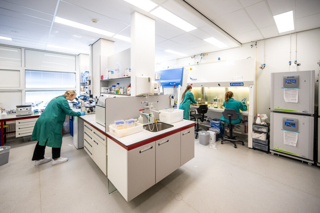
G-Cure and the UMCG division of experimental cardiology have extensive experience with the use of animal and human 3D cardiac models. With the use of these models we investigate the effects of (new) drugs on cardiac function in disease models, and examine the role of specific genes in the development of cardiovascular and metabolic diseases.
“Cardio Research has performed more than 500 phase 1, 2, and 3 clinical trials”
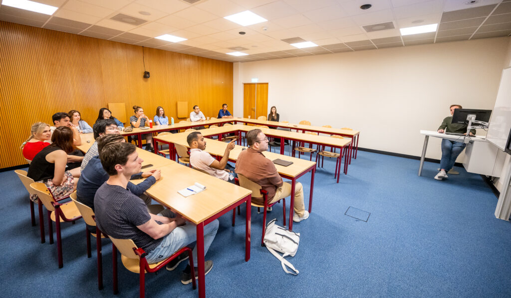
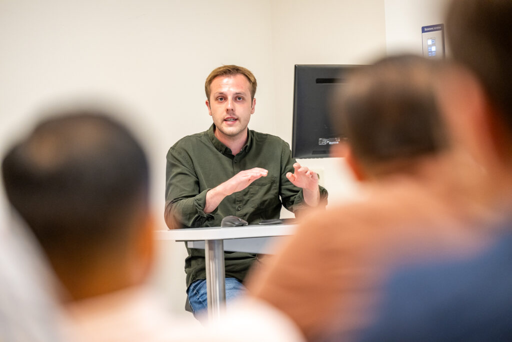
Since 1992, the department of cardiology has initiated a dedicated research department – Cardio Research. Cardio Research has performed more than 500 phase 1, 2, and 3 clinical trials in cardiology, both sponsored and investigator initiated trials. They have an outstanding record of patient recruitment.
Cardio Research employs project managers, research physicians, screening nurses, research nurses and research assistants.
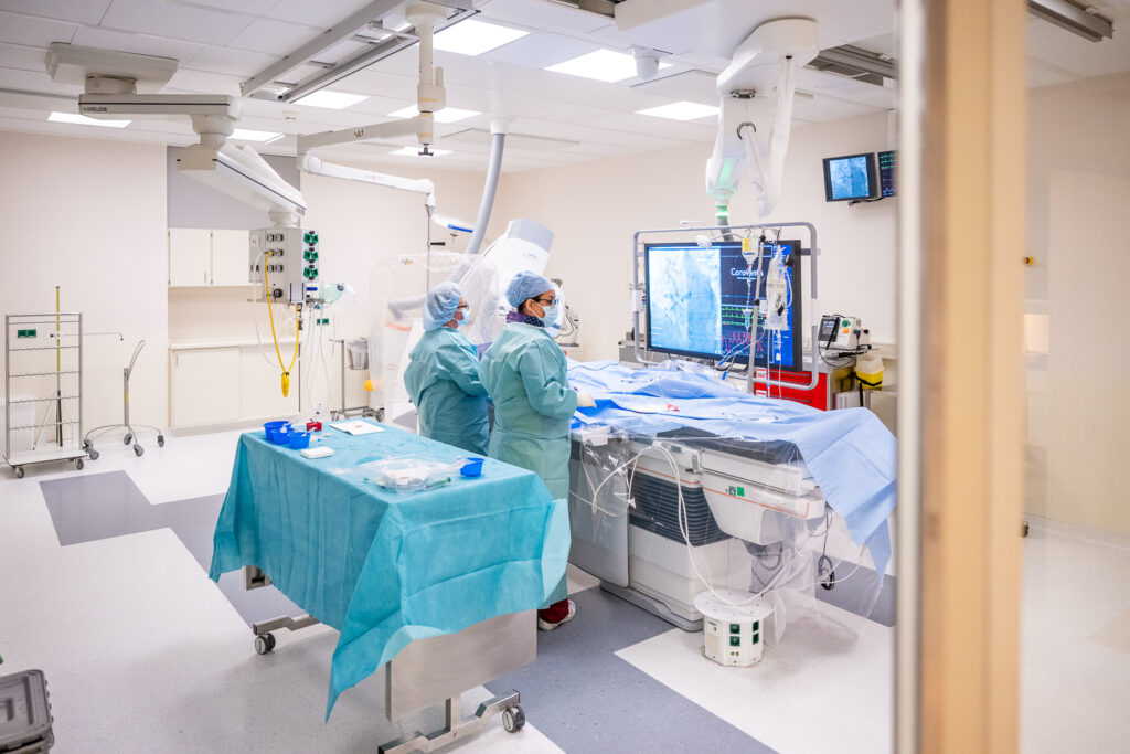
The department of cardiology employs 330 staff, including 30 cardiologists and 15 residents, and handles 5.300 hospital admissions and 18.000 outpatient visits per year. The Department of Cardiology conducts scientific research, with a special focus on heart failure, left ventricular remodeling, ischemic diseases, and atrial fibrillation. In addition, the department of cardiology initiated several studies on novel cardiovascular treatment options.